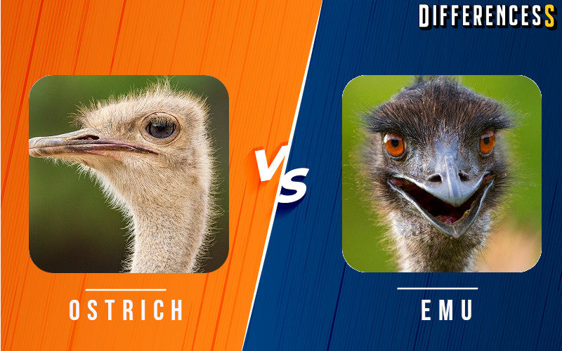
boy ultrasound vs girl ultrasound whats the difference 3115

Boy Ultrasound Vs Girl Ultrasound: What’s the Difference?

Ultrasound imaging is a process that uses sound waves to produce pictures of organs and body parts inside the body. Ultrasounds are commonly used for prenatal care, during childbirth, for diagnosing medical conditions, and for other purposes.

What is Ultrasound?

Ultrasound is a type of sound that travels through the air and is used to image objects inside the body. Ultrasound images are often used to diagnose medical conditions and help surgeons plan surgical procedures.
Ultrasound imaging is also used to create images of embryos before they are implanted in the mother’s womb. Boy vs girl ultrasound: What’s the difference?
There are some noticeable differences between boy and girl ultrasounds, but overall, they both provide valuable information about your baby. Here are four key points to keep in mind when deciding which type of ultrasound to have:

1) Girl ultrasounds typically use lower-frequency sound waves than boy ultrasounds, which allows them to image smaller structures more clearly.

2) Girl ultrasounds may be used to determine the sex of the baby early on in pregnancy, while boy ultrasounds may not be available until later in pregnancy.

3) Boy ultrasounds can provide images of the baby’s face and head, while girl ultrasounds are better at imaging the body and organs.

4) Both types of ultrasounds can be used during childbirth to help guide delivery.

Types of Ultrasounds

If you’re looking for a definitive answer to the question of “what’s the difference between boy ultrasound and girl ultrasound?” then you’re out of luck. Ultrasound is a medical procedure that uses sound waves to create images of body tissues and organs. So, while there are definite distinctions between male and female ultrasounds, they are by no means set in stone.

Generally speaking, male ultrasounds use higher-frequency sound waves to produce clearer images than female ultrasounds. Additionally, boy ultrasounds tend to be more detailed than girl ultrasounds, as they use slightly different frequency ranges. Finally, girl ultrasounds are often used during pregnancy to assess the development of the fetus. Boy ultrasounds are not generally used for this purpose.

So, if you’re looking for an all-encompassing answer to the question of “what’s the difference between boy ultrasound and girl ultrasound?” then you’ll be disappointed. However, if you’re simply interested in understanding the differences between these two types of ultrasounds, then you’re in good shape!

How Do Ultrasounds Work?

Ultrasounds are a type of medical imaging that uses sound waves to create images of parts of the body. They are used to diagnose problems and check for health conditions. Ultrasounds work differently for boys and girls. Here’s what you need to know about boy ultrasounds vs girl ultrasounds:

What is the difference between a boy ultrasound and a girl ultrasound?

The main difference between boy ultrasounds and girl ultrasounds is that boys typically have larger muscles and denser bones than girls, which can sometimes make it difficult to see certain areas of the body on a boy ultrasound. For this reason, some doctors may choose to perform a male ultrasound instead of a female ultrasound when they first assess a patient’s health.

Ultrasound Images and Their Uses

Ultrasound is a type of imaging that uses high-frequency sound waves to create pictures or videos of the inside of body tissues. Ultrasound images are commonly used in medicine to examine organs and tissues, as well as during pregnancy to check for problems such as fetal abnormalities. A common use of ultrasound is to determine the gender of a baby by looking at the structures inside the womb.

Ultrasound images are created by presenting a high-frequency sound wave to the surface of the skin and then capturing the image that is reflected back. The frequency of the sound waves determines how clearly you can see the image. Ultrasound machines use a number of different frequencies to create images that range from very detailed to providing an overall view of the body. Images can also be captured in multiple colors, which can help doctors determine details about tissue and organs.

The main difference between boy ultrasound and girl ultrasound is that boy ultrasound images typically have more detail than girl ultrasound images. This is because male babies typically have more prostate and testes than female babies, which are two common areas that need more detail during ultrasound imaging. Additionally, because boys tend to grow faster than girls, their organs may change size

What to Expect During an Ultrasound Exam

When you’re ready to schedule your ultrasound appointment, it’s important to understand the difference between a boy’s and girl’s ultrasound. Here’s what to expect during each exam.

A boy’s ultrasound is typically more detailed than a girl’s because there is more of the baby to see. Both exams provide information about the size and shape of the baby, but a boy’s ultrasound may also reveal problems such as an undescended testicle or cysts on the brain. A girl’s ultrasound may not show as many details, but it can still provide helpful information about the health of the baby.

Both exams are safe and can help you learn a lot about your baby. Just keep in mind that the results of a boy’s ultrasound may be different from those of a girl’s ultrasound. If you have questions about which exam is right for you, ask your doctor or ultrasound technician.

Boy vs Girl Ultrasounds: Differences and Similarities

Gender is one of the many things that can vary between boys and girls. Boys and girls also have different ultrasound features. Ultrasounds are a type of imaging that uses sound waves to create a picture of the inside of a body. Ultrasounds are generally used to look at babies, but they can also be used to look at other parts of the body, such as the brain and joints.

The main difference between boy ultrasounds and girl ultrasounds is that boy ultrasounds typically use higher-frequency sound waves than girl ultrasounds. This makes boy ultrasounds more detailed than girl ultrasounds, which is why they are sometimes used to look at baby’s brains and joints.

Another major difference between boy ultrasounds and girl ultrasounds is that girl ultrasounds tend to show more images of the internal organs, such as the uterus and ovaries. This is because the higher-frequency sound waves pass through these organs more easily than they do other parts of the body.

Overall, there are many similarities between boy ultrasounds and girl ultrasounds. Both types of ultrasound are very useful for getting a detailed image of what is happening inside a body. However, there are some important differences that

When to Have an Ultrasound

The gender of a baby is determined by the cells in the embryo’s earliest stage of development. For boys, these cells become testosterone producing cells, and for girls they become estrogen producing cells. This difference in hormone production can cause different physical characteristics in boys and girls.

Ultrasound is used to check the health of babies during their first few months of life. Ultrasound is not used to determine a baby’s gender. If you’re wondering whether to have an ultrasound scan for your baby, here are some things to keep in mind:

-Gender is determined by the cells in the embryo’s earliest stage of development. For boys, these cells become testosterone producing cells, and for girls they become estrogen producing cells. This difference in hormone production can cause different physical characteristics in boys and girls.

-The health of a baby is not always accurately reflected by ultrasound images. If you’re concerned about your baby’s health, ask your doctor about other tests that may be more accurate for detecting problems.

-Gender can be determined by a variety of methods other than ultrasound, including genetic testing or analysis of anatomy at birth. If you’re unsure whether you want to know your baby’s gender

What to Expect After an Ultrasound

When you go to the doctor for a ultrasound, you likely assume that the procedure will be the same for both men and women. However, there are some key differences between male and female ultrasounds that you should be aware of before your appointment. Here’s what to expect after an ultrasound for men and women:

1. Male Ultrasound Images Are Usually larger than Female Ultrasound Images
Many people assume that because ultrasound images are viewed on a computer or television screen, they will be the same size regardless of gender. This is not always the case. Male ultrasounds tend to be larger than female ultrasounds, which can result in more detailed images.

2. Male Ultrasounds May Provide More Information About Baby’s Health
Male ultrasounds can also provide more information about baby’s health than female ultrasounds. This is because male reproductive organs are located outside of the body, while female reproductive organs are located inside of the body.

3. It Can Be Difficult to Interpret Female Ultrasound Images
Female ultrasounds usually provide less information than male ultrasounds, making it difficult to interpret the images. In some cases, this may mean that doctors need to perform a

Male vs Female Anatomy

Ultrasound technology can be used to view the internal organs of both sexes, but there are some key anatomical differences between male and female bodies. In this blog, we’ll explore the main differences between male and female anatomy and how they impact ultrasounds.

When it comes to Ultrasound technology, males and females generally have the same images that can be seen with the use of this technology. However, there are a few key anatomical differences that must be taken into account when interpreting these images for each gender.

For example, when viewing images of the heart, the two chambers of the heart – left and right ventricles – will be clearly visible in a male image but may not be as easily seen in a female image. Additionally, because females have more blood flowing through their smaller veins than males do, certain areas ( like the neck) will appear more blue or green on ultrasound images than they would on an image of a male.

Despite these anatomical differences, Ultrasound scans are still an excellent way to view the internal organs of both genders. When choosing an Ultrasound provider, make sure to ask about their experience providing scans specifically for either gender, so you

Conclusion

Ultrasounds are a popular way to check on a baby’s development. Boys and girls both get ultrasounds during their pregnancies, but there are some key differences that you need to know about if you’re planning on having one. For example, boys tend to have more intense ultrasounds that can show problems with the baby’s heart or spine. On the other hand, girl ultrasounds often focus more on imaging the baby’s internal organs. So which is right for you? It really depends on your needs as a parent!

Similar Posts